Hetrotrophic Nutrition (For Classes 7 and 10) (NCERT page No 98 Class 10,Science Complete Explanation) ---PART 1---- By P K Verma
Class 7 Science NCERT Pg No 4-7
Hetrotrophic nutrition in which animals obtain organic food materials by consuming
bodies of products of other plants or animals are called
heterotrophs. Heterotrophic nutrition is of four types holozoic, saprozoic, parasitic and
symbiotic. In holozoic nutrition the solid food is ingested ,digested and then
absorbed into the cells. Animal maybe hervivores that is plant eaters for
example cow rabbit . They may be carnivorous that is flesh eaters for example dog
tiger.
They maybe Omnivores that is both plant and
animal eaters for example cockroach man.
They maybe insectivorous that is insect eaters like toads lizards.
They may be frugivorous that is Fruit
eaters for example monkeys Birds.
They may be sanguivores that is they feed on
blood of vertebrates for example female Anopheles Mosquito leeches.
They maybe
detrivores that is they feed on decaying organic matter for example
earthworms.
They may be fluid feeders that is the feed on Fluids from
plants for example butterflies male mosquitoes.
In Saprozoic nutrition organism obtain nutrients from
decaying organic materials after digesting the same with the help of enzymes. a few animals for example spider housefly secrete digestive enzymes directly
onto their food which are dead or decaying matters and then suck the digested
food.
In parasitic nutrition liquid food material is
obtained by the parasite from the body of host for example plasmodium,ascaris.
In symbiotic nutrition often two organism live in association with each other and derive nutrition from each other. For example Escherichia coli
in human intestine synthesizes vitamin B12 and in return obtained
simpler food from human intestine.
In holozoic nutrition involves in 4 steps.
These are
ingestion that is feeding of the food
Digestion that is breaking down of
complex organic molecules into simple molecules by hydrolysis.
Absorption
that is uptake of simple diffusible molecules by the digestive tract.
Egestion that is passing out of indigestible material through anus.
In different animals ingestion occurs by
different process on the basis of which they are classified into fluid feeder
animals, filter feeder animals and macrophages animals.
Ingestion occurs in
fluid feeder animals by diffusion for example parasitic protozoans
Pinocytosis
that is sanguivorous animals like leech mosquito etc.
Filter feeder animals are also called micropahagous animals are those animals which takes a small sized food particles.
Ingestion occurs in microphagous animals by maintaining a water current which
bring microscopic organism.examples are
Pseudopodial feeder that is Amoeba
Ciliary feeders that is Paramecium.
Flagellar feeders that is Sponges.
and Tentacular feeder that is hydra.
NCERT page 98 Class 10
Now Nutrition in Amoeba
Key Points
Protist cells may contain a single nucleus or many nuclei; they range in size from microscopic to thousands of meters in area.
Protists may have animal-like cell membranes, plant-like cell walls, or may be covered by a pellicle.
Some protists are heterotrophs and ingest food by phagocytosis, while other types of protists are photoautotrophs and store energy via photosynthesis.
Most protists are motile and generate movement with cilia, flagella, or pseudopodia.
Key Terms
amorphous : lacking a definite form or clear shape
multinucleate : having more than one nucleus
pellicle : cuticle, the hard protective outer layer of certain life forms
taxis : the movement of an organism in response to a stimulus; similar to kinesis, but more direct
phagocytosis : the process where a cell incorporates a particle by extending pseudopodia and drawing the particle into a vacuole of its cytoplasm
phagosome : a membrane-bound vacuole within a cell containing foreign material captured by phagocytosis.
Cell Structure
* The cells of protists are among the most elaborate and diverse of all cells.
* Most protists are microscopic and unicellular, but some true multicellular forms exist.
* A few protists live as colonies that behave in some ways as a group of free-living cells and in other ways as a multicellular organism.
* Single protist cells range in size from less than a micrometer to thousands of square meters (giant kelp).
* Animal-like cell membranes or plant-like cell walls envelope protist cells. In other protists, glassy silica-based shells or pellicles of interlocking protein strips encase the cells.
* The pellicle functions like a flexible coat of armor, preventing the protist from external damage without compromising its range of motion.
Metabolism
* Protists exhibit many forms of nutrition and may be aerobic or anaerobic.
* Protists that store energy by photosynthesis belong to a group of photoautotrophs and are characterized by the presence of chloroplasts.
* Other protists are heterotrophic and consume organic materials (such as other organisms) to obtain nutrition.
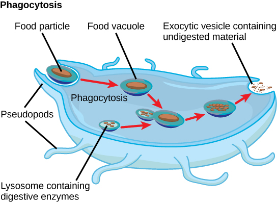
* Amoebas and some other heterotrophic protist species ingest particles by a process called phagocytosis in which the cell membrane engulfs a food particle and brings it inward, pinching off an intracellular membranous sac, or vesicle, called a food vacuole.
* The vesicle containing the ingested particle, the phagosome, then fuses with a lysosome containing hydrolytic enzymes to produce a phagolysosome, which breaks down the food particle into small molecules that diffuse into the cytoplasm for use in cellular metabolism.
* Undigested remains ultimately exit the cell via exocytosis.
Protist metabolism: The stages of phagocytosis include the engulfment of a food particle, the digestion of the particle using enzymes contained within a lysosome, and the expulsion of undigested materials from the cell.
* Subtypes of heterotrophs, called saprobes, absorb nutrients from dead organisms or their organic wastes.
* Some protists function as mixotrophs, obtaining nutrition by photoautotrophic or heterotrophic routes, depending on whether sunlight or organic nutrients are available.
Diet
Amoebas proteus eat algae, bacteria, plant cells, and microscopic protozoa and metazoa – some amoebas are parasites.
* They eat by surrounding tiny particles of food with pseudopods, forming a bubble-like food vacuole. The food vacuole digests the food.
* Wastes and excess water are transported outside the cell by contractile vacuoles.
* An amoeba, a single-celled organism lacking internal organs, approach a much smaller paramecium, which it begins to engulf with large outflowings of its cytoplasm, called pseudopodia.
* Once the paramecium is completely engulfed, a primitive digestive cavity, called a vacuole, forms around it.
* In the vacuole, acids break the paramecium down into chemicals that the amoeba can diffuse back into its cytoplasm for nourishment.
* Amoeba proteus captures a ciliate by surrounding it with its pseudopodia. The nucleus can be seen just below the pseudopodia. Bottom left is a water expelling contractile vacuole.
Amoebic Dysentry
Key Points
Amoebiasis is transmitted via food or water contaminated with fecal matter from an infected individual.
The pathogen, Entamoeba histolytica, is mainly found in tropical areas and is protected from degradation and destruction by its protective shell, formed during its cyst stage.
The cyst stage is typically passed in the feces and then ingested to cause infection.
The amoebas are able to burrow into the walls of the intestines, causing damage.
Key Terms
trophozoites : A protozoan in the feeding stage of its life cycle.
Amoebic dysentery, also referred to as amoebiasis, is caused by the ameoba -Entamoeba histolytica.
Dysentery is characterized as an inflammatory disorder of the intestine that results in severe diarrhea containing both mucus and blood in the feces, often accompanied with fever and abdominal pain.
The route of transmission for ameobic dysentery is the fecal-oral route.
Transmission and infection occur upon exposure or ingestion of contaminated food and water.
The infective cysts are passed via infected stool. The ameoba also demonstrates the ability to spread as free amoebae or trophozoites, meaning the cysts are not absolutely necessary; however, these states do not survive long outside of the host.
Sunday, March 29, 2020
Limiting Factors In Photosynthesis (For Class 7, 10 and 11)
* Law of minimum was proposed by a Libeg in 1843. * Law of limiting factors
was proposed by F.F Blackman in 1905. * The factor which is the minimum level is called the limiting factor .
when a process is conditioned as to its rapidity by a number of separate
factors the process is limited by the by the pace of slowest factor .This is called the law of limiting factor. * When the concentration of the limiting factor is increased to a certain
extent , the rate of process also increases. * Factors which influences the rate of photosynthesis are of two kinds. 1. External factors that is environmental factor and 2. Internal
factors. * The important environmental factors are number one light , carbon
dioxide concentration, oxygen concentration, temperature, water ,mineral salts. * The degradation of chlorophyll molecule due to high light intensity is
called photo oxidation. * Photo oxidation of chlorophyll under very high light
intensity is called solarization. * Rate of photosynthesis is maximum in red light and then in blue light. * Rate of photosynthesis is minimum in green light. * Rate of photosynthesis is more in intermittent light than in continuous
light. * If carbon dioxide concentration is increased from 0.03% to 1% the rate
of photosynthesis also increases. * When carbon dioxide concentration is more
than 1% the rate of photosynthesis decreases due to closure of stomata. * Carbon
dioxide reacts with water to form carbonic acid when it is in high
concentration. * The most common limiting factor for photosynthesis is carbon
dioxide. * Light occupies the next place. * Rate of
photosynthesis decreases in high concentration of oxygen. * The inhibitory effect
of oxygen on photosynthetic rate is called Warburg effect. * In C3 plants high
oxygen concentration leads to the occurrence of photorespiration as a result
net yield of photosynthesis decreases. * Temperature has maximum
influence on dark phase of photosynthesis. And hence the dark phase is called a thermochemical reaction. * Accumulation of Carbohydrates decreases the rate of
photosynthesis is due to non availability of space for new molecules. * As the
leaf expands the stomata , will be opened to the external atmosphere. Rate of
photosynthesis increases due to the entry of carbon dioxide.
Saturday, March 28, 2020
Photorespiration C3, C4 and CAM Plants (For Class 11) By PK Verma
All plants carry on photosynthesis by adding carbon dioxide (CO2) to a phosphorylated 5-carbon sugar called ribulose bisphosphate. This reaction is catalyzed by the enzyme ribulose bisphosphate carboxylase oxygenase (RUBISCO). The resulting 6-carbon compound breaks down into two molecules of 3-phosphoglyceric acid (PGA). These 3-carbon molecules serve as the starting material for the synthesis of glucose and other food molecules. The process is called the Calvin cycle and the pathway is called the C3 pathway.
C3 Cycle
Photorespiration
As its name suggests, RUBISCO catalyzes two different reactions:
adding CO2 to ribulose bisphosphate — the carboxylase activity
adding O2 to ribulose bisphosphate — the oxygenase activity
Which one predominates depends on the relative concentrations of O2 and CO2 with
high CO2, low O2 favoring the carboxylase action.
high O2, low CO2 favoring the oxygenase action.
The light reactions of photosynthesis liberate oxygen and more oxygen dissolves in the cytosol of the cell at higher temperatures. Therefore, high light intensities and high temperatures (above ~ 30°C) favor the second reaction.
The uptake of O2 by RUBISCO forms the 3-carbon molecule 3-phosphoglyceric acid, just as in the Calvin cycle, and the 2-carbon molecule glycolate. The glycolate enters peroxisomes where it uses O2 to form intermediates that enter mitochondria where they are broken down to CO2. So this process uses O2 and liberates CO2 as cellular respiration does which is why it is called photorespiration.
It undoes the good anabolic work of photosynthesis, reducing the net productivity of the plant. For this reason, much effort so far largely unsuccessful has gone into attempts to alter crop plants so that they carry on less photorespiration. The problem may solve itself. If atmospheric CO2 concentrations continue to rise, perhaps this will enhance the net productivity of the world's crops by reducing losses to photorespiration.
C4 Plants
Over 8,000 species of angiosperms have developed adaptations which minimize the losses to photorespiration. They all use a supplementary method of CO2 uptake which forms a 4-carbon molecule instead of the two 3-carbon molecules of the Calvin cycle. Hence these plants are called C4 plants. (Plants that have only the Calvin cycle are thus C3 plants). Some C4 plants - called CAM plants - separate their C3 and C4 cycles by time, while other C4 plants have structural changes in their leaf anatomy so that their C4 and C3 pathways are separated in different parts of the leaf with RUBISCO sequestered where the CO2 level is high; the O2 level low.
After entering through stomata, CO2 diffuses into a mesophyll cell. Being close to the leaf surface, these cells are exposed to high levels of O2, but they have no RUBISCO so cannot start photorespiration (nor the dark reactions of the Calvin cycle).
C4 Anatomy
Instead the CO2 is inserted into a 3-carbon compound (C3) called phosphoenolpyruvic acid (PEP) forming the 4-carbon compound oxaloacetic acid (C4).
Oxaloacetic acid is converted into malic acid or aspartic acid (both have 4 carbons), which is transported (by plasmodesmata) into a bundle sheath cell. Bundle sheath cells are deep in the leaf so atmospheric oxygen cannot diffuse easily to them and often have thylakoids with reduced photosystem II complexes (the one that produces O2). Both of these features keep oxygen levels low in Bundle sheath cells, which is where the 4-carbon compound is broken down into carbon dioxide, which enters the Calvin cycle to form sugars and starch, and pyruvic acid (C3), which is transported back to a mesophyll cell where it is converted back into PEP.
These C4 plants are well adapted to (and likely to be found in) habitats with high daytime temperatures and intense sunlight. Some examples crabgrass, corn (maize), sugarcane, and sorghum. Although only ~3% of the angiosperms, C4 plants are responsible for ~25% of all the photosynthesis on land.
C4 CELLS IN C3 PLANTS
The ability to use the C4 pathway has evolved repeatedly in different families of angiosperms - a remarkable example of convergent evolution. Perhaps the potential is in all angiosperms.
A report in the 24 January 2002 issue of Nature (by Julian M. Hibbard and W. Paul Quick) describes the discovery that tobacco, a C3 plant, has cells capable of fixing carbon dioxide by the C4 path. These cells are clustered around the veins (containing xylem and phloem) of the stems and also in the petioles of the leaves. In this location, they are far removed from the stomata that could provide atmospheric CO2. Instead, they get their CO2 and/or the 4-carbon malic acid in the sap that has been brought up in the xylem from the roots.
If this turns out to be true of many C3 plants, it would explain why it has been so easy for C4 plants to evolve from C3 ancestors.
CAM Plants
CAM plants are also C4 plants (CAM stands for crassulacean acid metabolism because it was first studied in members of the plant family Crassulaceae.). However, instead of segregating the C4 and C3 pathways in different parts of the leaf, CAM plants separate them in time instead (Table 1).
Comparision
Table 1
Night
Morning
CAM plants take in CO2 through their open stomata (they tend to have reduced numbers of them).
The CO2 joins with PEP to form the 4-carbon oxaloacetic acid.
This is converted to 4-carbon malic acid that accumulates during the night in the central vacuole of the cells.
The stomata close (thus conserving moisture as well as reducing the inward diffusion of oxygen).
The accumulated malic acid leaves the vacuole and is broken down to release CO2.
The CO2 is taken up into the Calvin (C3) cycle.
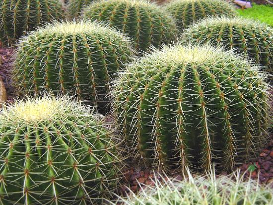
These adaptations also enable their owners to thrive in conditions of high daytime temperatures, intense sunlight, and low soil moisture. Some examples of CAM plants include cacti, Bryophyllum, the pineapple and all epiphytic bromeliads, sedums, and the "ice plant" that grows in sandy parts of the scrub forest biome.
Cultivated cacti in the Singapore Botanic Gardens.
Animation Video
Dark Reaction ( Calvin Cycle) For Class 10 and 11. By Pk Verma
Key Points
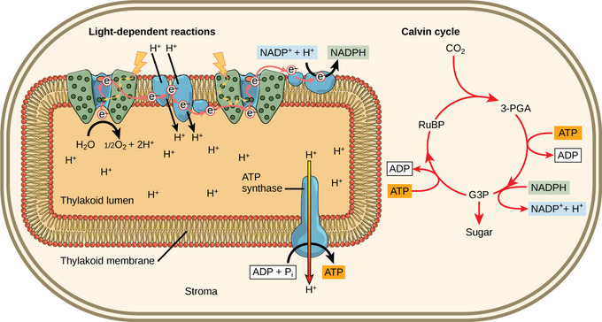
The Calvin cycle refers to the light-independent reactions in photosynthesis that take place in three key steps.
Although the Calvin Cycle is not directly dependent on light, it is indirectly dependent on light since the necessary energy carriers ( ATP and NADPH) are products of light-dependent reactions.
In fixation, the first stage of the Calvin cycle, light-independent reactions are initiated; CO2 is fixed from an inorganic to an organic molecule.
In the second stage, ATP and NADPH are used to reduce 3-PGA into G3P; then ATP and NADPH are converted to ADP and NADP+, respectively.
In the last stage of the Calvin Cycle, RuBP is regenerated, which enables the system to prepare for more CO2 to be fixed.
Key Terms
light-independent reaction: chemical reactions during photosynthesis that convert carbon dioxide and other compounds into glucose, taking place in the stroma
rubisco: (ribulose bisphosphate carboxylase) a plant enzyme which catalyzes the fixing of atmospheric carbon dioxide during photosynthesis by catalyzing the reaction between carbon dioxide and RuBP
ribulose bisphosphate: an organic substance that is involved in photosynthesis, reacts with carbon dioxide to form 3-PGA.
The Calvin Cycle
In plants, carbon dioxide (CO2) enters the leaves through stomata, where it diffuses over short distances through intercellular spaces until it reaches the mesophyll cells. Once in the mesophyll cells, CO2 diffuses into the stroma of the chloroplast, the site of light-independent reactions of photosynthesis. These reactions actually have several names associated with them. Other names for light-independent reactions include the Calvin cycle, the Calvin-Benson cycle, and dark reactions. The most outdated name is dark reactions, which can be misleading because it implies incorrectly that the reaction only occurs at night or is independent of light, which is why most scientists and instructors no longer use it.
Light Reactions: Light-dependent reactions harness energy from the sun to produce chemical bonds, ATP, and NADPH. These energy-carrying molecules are made in the stroma where the Calvin cycle takes place. The Calvin cycle is not totally independent of light since it relies on ATP and NADH, which are products of the light-dependent reactions.
The light-independent reactions of the Calvin cycle can be organized into three basic stages: fixation, reduction, and regeneration.
Stage 1: Fixation
In the stroma, in addition to CO2,two other components are present to initiate the light-independent reactions: an enzyme called ribulose bisphosphate carboxylase (RuBisCO) and three molecules of ribulose bisphosphate (RuBP). RuBP has five atoms of carbon, flanked by two phosphates. RuBisCO catalyzes a reaction between CO2 and RuBP. For each CO2 molecule that reacts with one RuBP, two molecules of 3-phosphoglyceric acid (3-PGA) form. 3-PGA has three carbons and one phosphate. Each turn of the cycle involves only one RuBP and one carbon dioxide and forms two molecules of 3-PGA. The number of carbon atoms remains the same, as the atoms move to form new bonds during the reactions (3 atoms from 3CO2 + 15 atoms from 3RuBP = 18 atoms in 3 atoms of 3-PGA). This process is called carbon fixation because CO2 is “fixed” from an inorganic form into organic molecules.
The Calvin Cycle: The Calvin cycle has three stages. In stage 1, the enzyme RuBisCO incorporates carbon dioxide into an organic molecule, 3-PGA. In stage 2, the organic molecule is reduced using electrons supplied by NADPH. In stage 3, RuBP, the molecule that starts the cycle, is regenerated so that the cycle can continue. Only one carbon dioxide molecule is incorporated at a time, so the cycle must be completed three times to produce a single three-carbon GA3P molecule, and six times to produce a six-carbon glucose molecule.
Stage 2: Reduction
ATP and NADPH are used to convert the six molecules of 3-PGA into six molecules of a chemical called glyceraldehyde 3-phosphate (G3P). This is a reduction reaction because it involves the gain of electrons by 3-PGA. Recall that a reduction is the gain of an electron by an atom or molecule. Six molecules of both ATP and NADPH are used. For ATP, energy is released with the loss of the terminal phosphate atom, converting it to ADP; for NADPH, both energy and a hydrogen atom are lost, converting it into NADP+. Both of these molecules return to the nearby light-dependent reactions to be reused and reenergized.
Stage 3: Regeneration
At this point, only one of the G3P molecules leaves the Calvin cycle and is sent to the cytoplasm to contribute to the formation of other compounds needed by the plant. Because the G3P exported from the chloroplast has three carbon atoms, it takes three “turns” of the Calvin cycle to fix enough net carbon to export one G3P. But each turn makes two G3Ps, thus three turns make six G3Ps. One is exported while the remaining five G3P molecules remain in the cycle and are used to regenerate RuBP, which enables the system to prepare for more CO2 to be fixed. Three more molecules of ATP are used in these regeneration reactions.


 C4 Anatomy